Файл:HeLa cell endocytic pathway labeled for EGFR and transferrin.jpg
Повна роздільність(3259 × 3510 пікселів, розмір файлу: 7,17 МБ, MIME-тип: image/jpeg)

Wikimedia Commons logo Відомості про цей файл містяться на Вікісховищі — централізованому сховищі вільних файлів мультимедіа для використання у проектах Фонду Вікімедіа.

Опис файлу

Опис
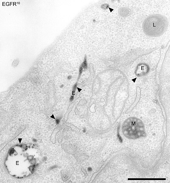
English: Compartments of the endocytic pathway in HeLa cells. Early endosomes (E), late endosomes/MVBs (M), and lysosomes (L) are visible. Epidermal growth factor receptors (EGFR) and transferrin (Tf) are labelled in the early endosomes.

Epidermal growth factor receptors are labelled with an antibody conjugated to 10 nm gold, applied to the cell surface. The cells were stimulated with EGF and allowed to internalize the receptors, bringing the gold with them. The image was taken 5 minutes after internalization, at which point most of the EGFR-gold has reached early endosomes (black dots, marked by arrowheads), but has not yet entered late endosomes or lysosomes. The cells are also loaded with transferrin conjugated to horseradish peroxidase (TfHRP). The HRP catalyses a reaction in the presence of DAB that produces a dark stain in the transferrin containing compartments in the image.

HeLa cells were preincubated for 1 h in serum-free medium. For the final 30 minutes the cells were incubated with TfHRP. The cells were then incubated with EGF and anti-EGFR 10 nm gold-conjugated antibody for 30 minutes at 4°C, washed, and incubated at 37°C for a further 5 minutes, all in the presence of TfHRP. Arrowheads; anti-EGFR 10 nm gold. Dark content; cross linked TfHRP. Bar, 500 nm.

Philips EM400 TEM

Methods for cell fixation and preparation for electron microscopy can be found in the reference below. Briefly, cells were room temperature fixed, a DAB reaction was done, and the cells were osmium stained, dehydrated and embedded en face in epon.

Related reference;

Doyotte, A., Russell, M.R.G., Hopkins, C.R., Woodman, P.G. (2005) Depletion of TSG101 forms a mammalian ‘‘Class E’’ compartment: a multicisternal early endosome with multiple sorting defects. J. Cell Sci, 118:3003-3017.
Час створення
Джерело Власна робота
Автор Matthew R G Russell

Ліцензування

Я, власник авторських прав на цей твір, добровільно публікую його на умовах таких ліцензій:
GNU head Дозволяється копіювати, розповсюджувати та/або модифікувати цей документ на умовах ліцензії GNU FDL версії 1.2 або більш пізньої, виданої Фондом вільного програмного забезпечення, без незмінних розділів, без текстів, які розміщені на першій та останній обкладинці. Копія ліцензії знаходиться у розділі GNU Free Documentation License.
w:uk:Creative Commons
зазначення авторства
Цей файл ліцензований на умовах Creative Commons Attribution 3.0 Unported
Ви можете вільно:
  • ділитися – копіювати, поширювати і передавати твір
  • модифікувати – переробляти твір
При дотриманні таких умов:
  • зазначення авторства – Ви повинні вказати авторство, надати посилання на ліцензію і вказати, чи якісь зміни було внесено до оригінального твору. Ви можете зробити це в будь-який розсудливий спосіб, але так, щоб він жодним чином не натякав на те, наче ліцензіар підтримує Вас чи Ваш спосіб використання твору.
Ви можете обрати ліцензію на ваш розсуд.

Підписи

Додайте однорядкове пояснення, що саме репрезентує цей файл

Об'єкти, показані на цьому файлі

зображує

Історія файлу

Клацніть на дату/час, щоб переглянути, як тоді виглядав файл.

Дата/часМініатюраРозмір об'єктаКористувачКоментар
поточний22:22, 3 листопада 2009Мініатюра для версії від 22:22, 3 листопада 20093259 × 3510 (7,17 МБ)Putneybridgetube{{Information |Description={{en|1='''Compartments of the endocytic pathway in human HeLa cells.''' Early endosomes (E), late endosomes/MVBs (M), and lysosomes (L) are visible. Epidermal growth factor receptors (EGFR) and transferrin (Tf) are labelled in

Така сторінка використовує цей файл:

Глобальне використання файлу

Цей файл використовують такі інші вікі:

Метадані