Файл:Brain - Lobes.png
Нема версії з більшою роздільністю.
Brain_-_Lobes.png (701 × 487 пікселів, розмір файлу: 360 КБ, MIME-тип: image/png)
| Відомості про цей файл містяться на Вікісховищі — централізованому сховищі вільних файлів мультимедіа для використання у проектах Фонду Вікімедіа. |
| Опис |
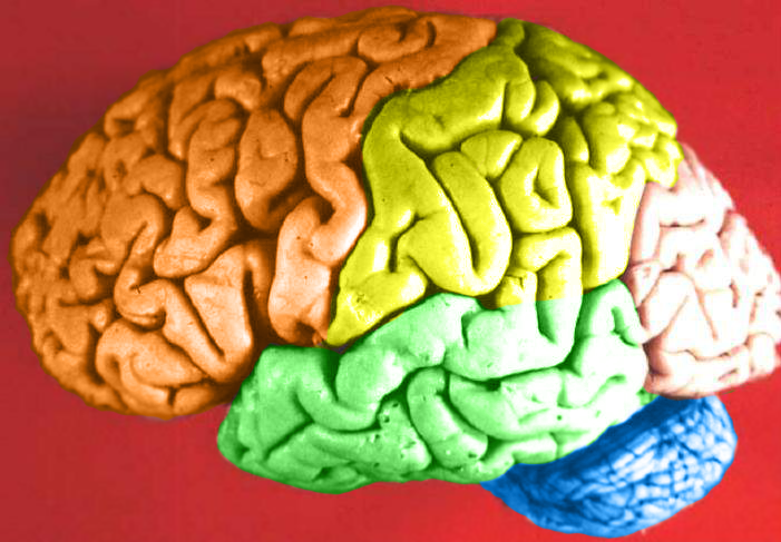
Human brain lateral view - Lobes
|
| Час створення | (UTC) |
| Джерело | Human_brain_lateral_view_description.JPG |
| Автор | Dep't. of Cellular Biology & Anatomy, Louisiana State University Health Sciences Center Shreveport |
| Ліцензія (Повторне використання цього файлу) |
CC-BY |
| Інші версії |
| Це відредаговане зображення, тобто одержане цифровою обробкою з оригіналу. Зміни: Hemispheres in color.. Початкове зображення знаходиться тут: Human brain lateral view description.JPG. Відредагував DavoO.
|
Ліцензування
Я, власник авторських прав на цей твір, добровільно публікую його на умовах такої ліцензії:
Цей файл доступний на умовах ліцензії Creative Commons Attribution 2.5 Generic
- Ви можете вільно:
- ділитися – копіювати, поширювати і передавати твір
- модифікувати – переробляти твір
- При дотриманні таких умов:
- зазначення авторства – Ви повинні вказати авторство, надати посилання на ліцензію і вказати, чи якісь зміни було внесено до оригінального твору. Ви можете зробити це в будь-який розсудливий спосіб, але так, щоб він жодним чином не натякав на те, наче ліцензіар підтримує Вас чи Ваш спосіб використання твору.
The following refers to the original source file, not this derivative version.
Цей файл, був спочатку опублікований на зовнішньому сайті https://web.archive.org/web/20110514023714/http://www.healcentral.org/healapp/showMetadata?metadataId=40566, був перевірений 1 листопада 2013 перевіряючим Avenue, який підтвердив, що у ту дату файл був там доступний на умовах зазначеної ліцензії.
|
Журнал завантажень локального файлу
This image is a derivative work of the following images:
- File:Human_brain_lateral_view_description.JPG licensed with Cc-by-2.5
- 2006-06-20T13:58:22Z Patho 701x487 (50176 Bytes) {{Information| |Description='''Human brain lateral view - Lobes''' # Lobus frontalis # Lobus parietalis # Lobus temporalis # Lobus occipitalis # Sulcus lateralis # Sulcus centralis # Sulcus parietooccipitalis # Incisura preo
- 2006-06-20T13:54:13Z Patho 701x487 (49891 Bytes) Auf eine alte Version zurückgesetzt
- 2006-06-20T13:51:38Z Patho 701x487 (50074 Bytes) {{Information| |Description='''Human brain lateral view - Lobes''' # Lobus frontalis # Lobus parietalis # Lobus temporalis # Lobus occipitalis # Sulcus lateralis # Sulcus centralis # Sulcus parietooccipitalis # Incisura preo
- 2006-06-20T13:28:44Z Patho 701x487 (49891 Bytes) {{Information| |Description='''Human brain lateral view''' # Lobus frontalis # Lobus parietalis # Lobus temporalis # Lobus occipitalis # sulcus lateralis # Sulcis centralis # Sulcus parietooccipitalis # Incisura preoccipital
Uploaded with derivativeFX
Історія файлу
Клацніть на дату/час, щоб переглянути, як тоді виглядав файл.
| Дата/час | Мініатюра | Розмір об'єкта | Користувач | Коментар | |
|---|---|---|---|---|---|
| поточний | 22:11, 23 лютого 2009 | | 701 × 487 (360 КБ) | DavoO | {{Information |Description='''Human brain lateral view - Lobes''' # Lobus frontalis # Lobus parietalis # Lobus temporalis # Lobus occipitalis # Sulcus lateralis # Sulcus centralis # Sulcus parietooccipitalis # Incisura preoccipitalis # Polus frontalis # |
Використання файлу
Нема сторінок, що використовують цей файл.
Глобальне використання файлу
Цей файл використовують такі інші вікі:
- Використання в cs.wikipedia.org
- Використання в en.wikipedia.org
- Talk:Alcohol intoxication
- Talk:LSD
- Talk:Scopolamine
- Talk:Qigong
- Talk:Recreational drug use
- Talk:Psilocybin
- Talk:Phenomenology (philosophy)
- Talk:Alcohol (chemistry)
- Talk:Timothy Leary
- Talk:Psilocybe cubensis
- Talk:Nitrous oxide
- Talk:Atropine
- Talk:Out-of-body experience
- Talk:The Doors of Perception
- Talk:Carlos Castaneda
- Talk:Ganzfeld experiment
- Talk:Meditation
- Talk:Zen
- Talk:Hysteria
- Talk:Peyote
- Talk:Hashish
- Talk:Ketamine
- Talk:Coca
- Talk:Hippie
- Talk:Spirituality
- Talk:Mantra
- Talk:N,N-Dimethyltryptamine
- Talk:Amanita muscaria
- Talk:Hookah
- Talk:Autogenic training
- Talk:Psychonautics
- Talk:Hypnosis
- Talk:Dipropyltryptamine
- Talk:Ergot
- Talk:Phencyclidine
- Talk:Anadenanthera peregrina
- Talk:Shamanism
- Talk:Mescaline
- Talk:Diphenhydramine
- Talk:Salvinorin A
- Talk:Human Potential Movement
- Talk:Argyreia nervosa
- Talk:Terence McKenna
- Talk:Psilocybin mushroom
- Talk:Atropa belladonna
- Talk:Hypnagogia
- Talk:Kundalini yoga
- Talk:DiPT
- Talk:Dreamachine
Переглянути сторінку глобального використання цього файлу.