Файл:Surface projections of the organs of the trunk.png

Повна роздільність(1583 × 2533 пікселів, розмір файлу: 3,33 МБ, MIME-тип: image/png)

Wikimedia Commons logo Відомості про цей файл містяться на Вікісховищі — централізованому сховищі вільних файлів мультимедіа для використання у проектах Фонду Вікімедіа.

Опис файлу

Опис
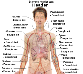
English: Surface projections of the major organs of the trunk, using the vertebral column and rib cage as main reference points of superficial anatomy. The transpyloric plane and McBurney's point are among the marked locations.

To discuss image, please see Talk:Human body diagrams

Час створення
Джерело

All included images are in public domain. See Template:Human body diagrams for individual details.

References

Spinal vertebrae levels:

  • Located at middle of frontal face of spinal vertebrae according to reference the image :File:Gray 111 - Vertebral column.png when pasted into image to superiorly fit the surface bulges of the clavicular bones and inferiorly fit the surface bulges of the anterior iliac spine.

Gross overview of organ locations:

  • Plate 260B - Regions and Planes of Adbomen. From: The Interactive Atlas of Human Anatomy Version 3.0 (CD-ROM). Authors: Frank H. Netter and Carlos A. G. Machado. Published by Saunders, October 1, 2002. ISBN-10: 1929007140. ISBN-13: 978-1929007141.
  • Fig 139 from The SPENCER SYSTEM. SEVENTEENTH EDITION
  • Plate 1034 from Gray's Anatomy

Locations of specific organs:
The following vertebral levels are generally given by the middle of the vertebral body.

  • Bålens ytanatomy (Superficial anatomy of the trunk). Anca Dragomir, Mats Hjortberg and Godfried M. Romans. Section for human anatomy at the Department of medical cell biology, Uppsala university, Sweden.
Key points from that compendium:
  • The oblique fissure of the right lung goes from the intercostal space 4 to 6
  • The horizontal fissure goes along the 4th rib (the original image from Gray has this line almost at the 5th rib, but this is not labeled in image, as other lung images seem to support the upper level)
  • The cardia of the stomach is at the level of the 10th rib
  • The fundus of the stomach is in the 5th intercostal space, slightly below the apex of the heart
  • Pylorus is usually at the L1 (the transpyloric plane)
  • The transpyloric plane is at the level of:
    • lumbar vertebra 1
    • the pancreatic body
    • the origins of the superior mesenteric artery from the aorta and portal vein
    • the left and right colic flexure
    • hilum of the kidney on the left
    • upper pole of the kidney on the right
    • duodenojejunal flexure
  • McBurney's point is located one third of a line from the anterior superior iliac spine and the navel
  • The upper rim of the liver is in the 5th intercostal space
  • The left lobe of the liver is to the left of the midline
  • The lower limit of the liver at the midline is approximately 3 cm below processus xiphoideus
  • The spleen is projected against the 9th and 10th rib on the backside
  • The left kidney is approximately at the vertebral level T12 to L3, and the right one slightly lower.
  • Atlas of Human Anatomy in Cross Section: Appendix: Topography of the Thorax and Abdomen. Ronald A. Bergman, Ph.D., Adel K. Afifi, M.D., Jean J. Jew, M.D., and Paul C. Reimann, B.S. [1]:
  • The average position of the highest point of the spleen is at the level of the lower third of the tenth thoracic vertebra.
  • The average position of the lowest point of the spleen is at the level of the middle third of the first lumbar vertebra.

Position of the navel

Urinary tract:

Автор Mikael Häggström
Є векторний варіант цього зображення (SVG).
Його слід використовувати замість цієї растрової версії для більш якісного відображення.

File:Surface projections of the organs of the trunk.png → File:Surface projections of the organs of the trunk.svg

Щоб дізнатися більше про векторну графіку, прочитайте статтю «Перетворення зображень у формат SVG».
Також доступна інформація про підтримку формату SVG у MediaWiki.

На інших мовах
Alemannisch  Bahasa Indonesia  Bahasa Melayu  British English  català  čeština  dansk  Deutsch  eesti  English  español  Esperanto  euskara  français  Frysk  galego  hrvatski  Ido  italiano  lietuvių  magyar  Nederlands  norsk bokmål  norsk nynorsk  occitan  Plattdüütsch  polski  português  português do Brasil  română  Scots  sicilianu  slovenčina  slovenščina  suomi  svenska  Tiếng Việt  Türkçe  vèneto  Ελληνικά  беларуская (тарашкевіца)  български  македонски  нохчийн  русский  српски / srpski  татарча/tatarça  українська  ქართული  հայերեն  বাংলা  தமிழ்  മലയാളം  ไทย  한국어  日本語  简体中文  繁體中文  עברית  العربية  فارسی  +/−
Нове зображення

Ліцензування

Public domain Я, власник авторських прав на цю роботу, передаю роботу в суспільне надбання. Застосовується по всьому світу.
У деяких країнах це не може бути юридично можливо, в такому випадку:
Я даю кожному право на використання цієї роботи для будь-яких цілей, без будь-яких умов, якщо такі умови не вимагаються за законом.

Human body diagrams

Main article at: Human body diagrams

Template location:Template:Human body diagrams

How to derive an image

Derive directly from raster image with organs

The raster (.png format) images below have most commonly used organs already included, and text and lines can be added in almost any graphics editor. This is the easiest method, but does not leave any room for customizing what organs are shown.

Adding text and lines:

Derive "from scratch"

By this method, body diagrams can be derived by pasting organs into one of the "plain" body images shown below. This method requires a graphics editor that can handle transparent images, in order to avoid white squares around the organs when pasting onto the body image. Pictures of organs are found on the project's main page. These were originally adapted to fit the male shadow/silhouette.

Organs:

More organs are found at: Human body diagrams/Organs

Derive by vector template

The Vector templates below can be used to derive images with, for example, Inkscape. This is the method with the greatest potential.

See Human body diagrams/Inkscape tutorial for a basic description in how to do this.

Examples of derived works

More examples

Licensing

Creative Commons CC-Zero Цей файл доступний на умовах Creative Commons CC0 1.0 Universal Public Domain Dedication.
Особа, що пов'язала роботу з даною дією, передала роботу у суспільне надбання шляхом відмови від усіх своїх прав на роботу по всьому світу по закону про авторське право, включаючи всі пов'язані і суміжні права, в тій мірі, що допускається законом.

Ви можете копіювати, змінювати, розповсюджувати і виконувати роботу, навіть на комерційній основі, не питаючи дозволу.

Підписи

Додайте однорядкове пояснення, що саме репрезентує цей файл

Об'єкти, показані на цьому файлі

зображує

Історія файлу

Клацніть на дату/час, щоб переглянути, як тоді виглядав файл.

(найновіші | найдавніші) Переглянути (новіших 10 | ) (10 | 20 | 50 | 100 | 250 | 500).
Дата/часМініатюраРозмір об'єктаКористувачКоментар
поточний09:19, 27 грудня 2019Мініатюра для версії від 09:19, 27 грудня 20191583 × 2533 (3,33 МБ)Mikael Häggström+Costal margin
10:38, 11 листопада 2010Мініатюра для версії від 10:38, 11 листопада 20101050 × 1680 (2,07 МБ)Mikael HäggströmAdapted to recently added overview images. Distinguished different ways to designate vertebrae levels.
10:04, 7 листопада 2010Мініатюра для версії від 10:04, 7 листопада 2010936 × 1325 (1,77 МБ)Mikael Häggströmupdate from svg
09:46, 7 листопада 2010Мініатюра для версії від 09:46, 7 листопада 2010936 × 1325 (1,77 МБ)Mikael Häggströmupdate from svg
04:51, 24 жовтня 2010Мініатюра для версії від 04:51, 24 жовтня 2010936 × 1325 (1,61 МБ)Mikael HäggströmSmoother edges
05:18, 10 жовтня 2010Мініатюра для версії від 05:18, 10 жовтня 2010936 × 1325 (1,61 МБ)Mikael HäggströmMinor kidney adjustment. More realistic hip bone
04:47, 6 жовтня 2010Мініатюра для версії від 04:47, 6 жовтня 2010936 × 1325 (1,73 МБ)Mikael HäggströmDistinguished stomach and spleen. Removed painted arteries out of scope.
18:40, 4 жовтня 2010Мініатюра для версії від 18:40, 4 жовтня 2010936 × 1325 (1,74 МБ)Mikael HäggströmLowered spleen
15:21, 3 жовтня 2010Мініатюра для версії від 15:21, 3 жовтня 2010936 × 1325 (1,74 МБ)Mikael HäggströmDecreased some opacity. Aligned tail of pancreas with spleen. Adjusted fissure marking width.
18:20, 2 жовтня 2010Мініатюра для версії від 18:20, 2 жовтня 2010936 × 1325 (1,72 МБ)Mikael Häggström+liver label
(найновіші | найдавніші) Переглянути (новіших 10 | ) (10 | 20 | 50 | 100 | 250 | 500).

Така сторінка використовує цей файл:

Глобальне використання файлу

Цей файл використовують такі інші вікі:

Переглянути сторінку глобального використання цього файлу.

Метадані